Advanced Catheter-Based Treatment for Deep Vein Thrombosis
This article explains catheter-guided thrombolysis as a safe and minimally invasive treatment for deep vein thrombosis. It covers the procedure, benefits, preparation tips, equipment used, and potential risks involved, providing a comprehensive overview for patients and healthcare providers seeking effective DVT management options.
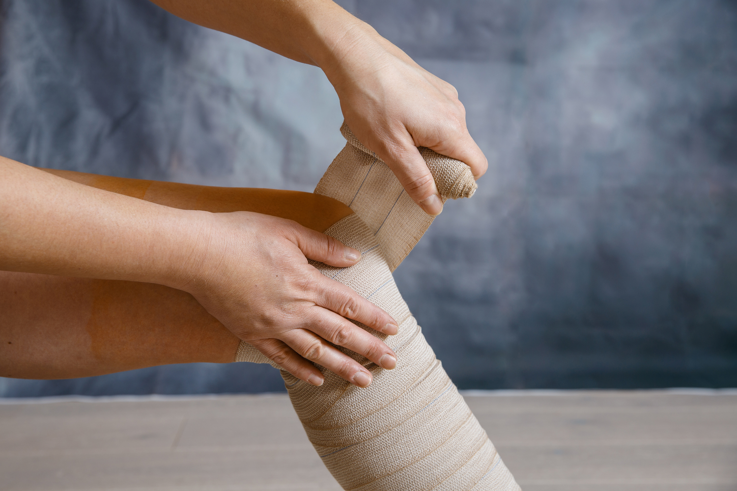
Overview of minimally invasive thrombolytic procedures
Deep vein thrombosis (DVT) involves clot formation in deep veins, typically in the legs or thighs. Symptoms include redness, swelling, and tenderness along the affected area. It predominantly affects individuals over 50 or those with impaired circulation due to underlying conditions.
What is catheter-guided thrombolysis?
This technique employs X-ray-guided catheters to dissolve clots within veins.
It helps restore normal blood flow, preventing tissue damage caused by blockages.
Benefits of this approach
A safe, minimally invasive option for DVT management.
Improves circulation without the need for open surgery.
No large incisions are required, resulting in shorter recovery periods.
Patients often avoid prolonged hospital stays typical of traditional surgery.
Pre-treatment preparation tips
Inform your doctor about all current and past medications.
Disclose any existing health conditions.
If pregnant or planning pregnancy, make sure your healthcare provider is aware.
Blood tests may be performed to evaluate kidney function and clotting status.
Your physician might recommend medication adjustments or preparatory steps.
Procedure steps
Identify the target vein using contrast dye and X-ray guidance.
Create a small skin puncture to insert the catheter.
Advance the catheter to the site of the clot.
Use medication or devices to break up or dissolve the clot.
Needed equipment
A thin plastic catheter similar in size to spaghetti.
X-ray machines, medication delivery systems, monitors, ultrasound tools, and IV lines.
Specific tools may vary based on your doctor's treatment plan.
Evaluating treatment outcomes
An interventional radiologist reviews the success of the procedure.
Follow-up treatments may be necessary if there was tissue damage.
Additional testing such as blood work and imaging might be performed during follow-up visits.
Possible risks and considerations
Infection risk is approximately 0.1%.
Rare allergic reactions or injuries to vessels.
Bleeding complications at various sites.
Patients with kidney problems could experience rare kidney damage.
Clot fragments may migrate, requiring further intervention.