Comprehensive Guide to Bone Density Tests and Why They Matter
Discover essential information about bone density testing, its role in diagnosing osteoporosis, and how it helps evaluate fracture risk. The non-invasive scan assesses bones' mineral density using low-dose X-rays, focusing on hips, spine, and other key areas. Whether monitoring treatment or screening at-risk individuals, this quick test provides vital insights into bone health, particularly for postmenopausal women and those with risk factors. Early detection can prevent fractures and support effective management of osteoporosis.
Comprehensive Guide to Bone Density Tests and Why They Matter
Osteoporosis is a condition that causes bones to become fragile, increasing fracture risk, particularly in the hips, spine, and wrists. While women are more frequently affected, men can also develop this condition. In the United States, around 10 million people—mainly women—are diagnosed, with approximately 34 million at risk due to low bone mass, known as osteopenia.
A bone density assessment is a swift, non-invasive test that screens for osteoporosis and helps track treatment progress.
Doctors may suggest a bone density exam if:
Height loss: A drop of 1.6 inches or more could signal spinal fractures related to osteoporosis.
Fractures: Fragile bones tend to break more easily.
Medication: Long-term use of steroids like prednisone can impair bone healing, increasing osteoporosis risk.
Organ transplants: Recipients are more prone to bone issues due to immunosuppressant effects.
Hormonal shifts: Postmenopausal women experience decreased estrogen, and cancer treatments can reduce hormones such as testosterone in men, elevating osteoporosis risk.
The procedure uses a low-dose X-ray to detect bone mineral loss, mainly targeting the hips and spine. If hip scans aren't possible, forearm scans are performed. For those under 60, hip assessments are generally preferred.
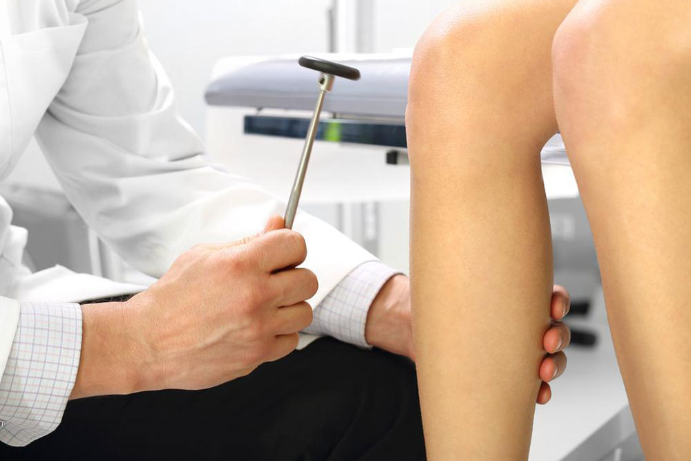
Whole-body, spine, and hip assessments are done with central machines, while peripheral devices examine the heel, shinbone, wrist, knee, and fingers. The test takes about 15 minutes, with the person lying down for 5-8 minutes. 3D imaging provides detailed insights into age and disease-related bone changes beyond osteoporosis.